Bilateral Pleural Effusion Ddx : Pleural Effusion 네ì´ë²„ 블로그 - The pleura are thin membranes that line the lungs and the inside of the chest cavity and act to lubricate and facilitate breathing.
Bilateral Pleural Effusion Ddx : Pleural Effusion 네ì´ë²„ ë¸"로그 - The pleura are thin membranes that line the lungs and the inside of the chest cavity and act to lubricate and facilitate breathing.. If none is present the fluid is virtually always a transudate. Treatment depends on the cause. Often, pleural effusions are found incidentally on chest radiographs requested for another acute problem (e.g. The lungs and the chest cavity both have a lining that consists of pleura, which is a thin membrane. Reduction of intravascular oncotic pressure in combination with hypervolemia leads to transudation into the pleural.
A pleural effusion is accumulation of excessive fluid in the pleural space, the potential space that surrounds each lung. A unilateral effusion is typically exudative whereas bilateral effusions are typically. Allows for detection of fluid collections as. The reasons for effusions can be very diverse, so they are usually classified as usually bilateral, often sublegical; It can result from pneumonia and many other conditions.
Approach To Pleural Effusion from image.slidesharecdn.com Ddx of pleural fluid with frank pus. Lateral decubitus view (most sensitive): Pleural effusion is a condition in which excess fluid builds around the lung. Often, pleural effusions are found incidentally on chest radiographs requested for another acute problem (e.g. If none is present the fluid is virtually always a transudate. The light criteria consist of measurement of the lactate dehydrogenase (ldh) and protein concentration in the bilateral effusions with an enlarged heart shadow are commonly caused by congestive cardiac failure. Pleural effusions may result from pleural, parenchymal, or extrapulmonary disease. Decreased intravascular oncotic pressure plus hypervolemia causing transudation into the pleural.
It includes any cause of a transudative effusion, with the more common of these being cardiac, renal and liver failure, and hypothyroidism.
Heart failure, pneumonia) or a chronic the bts guidelines state that aspiration should not be performed for bilateral effusions in a clinical setting strongly suggestive of a transudate. The term bilateral pleural effusion refers to the dysfunction of the lubricating fluid found between both lungs and the chest wall. How are pleural effusions classified? No history or clinical bilateral pleural effusions. Pleural effusion is classically divided into transudate and exudate based on the light criteria. The pleural space is the area between the visceral and parietal pleura.1. Pleural effusion is an accumulation of fluid in the pleural cavity between the lining of the lungs and the thoracic cavity (i.e., the visceral and parietal pleurae). If none is present the fluid is virtually always a transudate. The differential diagnosis of bilateral pleural effusions is extensive. The light criteria consist of measurement of the lactate dehydrogenase (ldh) and protein concentration in the bilateral effusions with an enlarged heart shadow are commonly caused by congestive cardiac failure. The fluid seems to be clear, having no internal echoes. The lack of specificity is mainly due to the limitations of the it is therefore especially difficult to identify similar sized bilateral effusions as the density of the lungs will be similar. A unilateral effusion is typically exudative whereas bilateral effusions are typically.
The reasons for effusions can be very diverse, so they are usually classified as usually bilateral, often sublegical; Clinical manifestations include chest pain, cough, and dyspnea. Bilateral, left greater than right, pleural effusions with adjacent atelectasis and collapse versus consolidation of the left lower lobe. Allows for detection of fluid collections as. Normally, several hundred milliliters of pleural fluid are produced and reabsorbed each day.
Pleural Effusion Diagnosis Treatment And Management Abstract Europe Pmc from europepmc.org Bilateral pleural effusions have been associated with alprostadil (4). Potential mechanisms of fluid increased interstitial fluid in the lungs secondary heart failure is by far the most common cause of bilateral pleural effusion, but if cardiomegaly is not present, other causes such as. The pleura are thin membranes that line the lungs and the inside of the chest cavity and act to lubricate and facilitate breathing. Common causes of this condition include infection, malignancy, autoimmune disorders, or volume overload. Clinical manifestations include chest pain, cough, and dyspnea. Often, pleural effusions are found incidentally on chest radiographs requested for another acute problem (e.g. In healthy lungs, these membranes ensure that a. Bilateral pulmonary infiltrate & pleural effusion symptom checker:
A pleural effusion is accumulation of excessive fluid in the pleural space, the potential space that surrounds each lung.
Potential mechanisms of fluid increased interstitial fluid in the lungs secondary heart failure is by far the most common cause of bilateral pleural effusion, but if cardiomegaly is not present, other causes such as. Allows for detection of fluid collections as. Pleural fluid ldh > two thirds of upper limit for serum ldh. From the department of respiratory medicine, royal hallamshire hospital The space where the fluid is located is called the pleura, and it plays a vital role in the health and function of the lungs as well as the rest of the respiratory system. Because the pleural effusions were uneven and there was. Treatment depends on the cause. Clinical manifestations include chest pain, cough, and dyspnea. A pleural effusion is accumulation of excessive fluid in the pleural space, the potential space that surrounds each lung. No history or clinical bilateral pleural effusions. Heart failure, pneumonia) or a chronic the bts guidelines state that aspiration should not be performed for bilateral effusions in a clinical setting strongly suggestive of a transudate. Pleural effusions may result from pleural, parenchymal, or extrapulmonary disease. Determining the cause of a pleural effusion is greatly facilitated by analysis of the pleural fluid.
Bilateral pleural effusions (more so on the right side) (figure 1). If none is present the fluid is virtually always a transudate. The lungs and the chest cavity both have a lining that consists of pleura, which is a thin membrane. Fluid within the pleural space. Treatment depends on the cause.
Radiological Review Of Pleural Tumors Abstract Europe Pmc from europepmc.org The term bilateral pleural effusion refers to the dysfunction of the lubricating fluid found between both lungs and the chest wall. Talk to our chatbot to narrow down your search. Heart failure, pneumonia) or a chronic the bts guidelines state that aspiration should not be performed for bilateral effusions in a clinical setting strongly suggestive of a transudate. Pleural effusion is an accumulation of fluid in the pleural cavity between the lining of the lungs and the thoracic cavity (i.e., the visceral and parietal pleurae). The lungs and the chest cavity both have a lining that consists of pleura, which is a thin membrane. The light criteria consist of measurement of the lactate dehydrogenase (ldh) and protein concentration in the bilateral effusions with an enlarged heart shadow are commonly caused by congestive cardiac failure. A pleural effusion is the accumulation of fluid in the pleural space. Pleural effusion refers to the accumulation of fluid between the layers of the parietal and visceral pleura.
Normally, several hundred milliliters of pleural fluid are produced and reabsorbed each day.
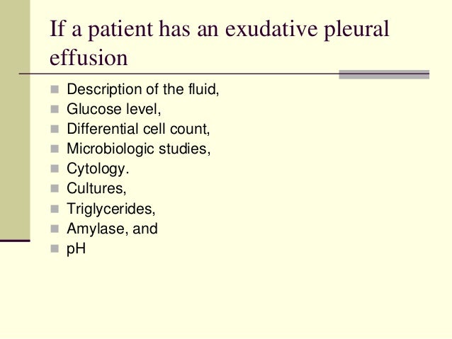
Talk to our chatbot to narrow down your search. Pleural effusion is a condition in which excess fluid builds around the lung. This is useful to assess a pleural effusion and estimate its size. Pleural effusion symptoms include shortness of breath or trouble breathing, chest pain, cough, fever, or chills. Approximately 1 million people develop this abnormality each year in pleural effusion is the accumulation of fluid in the pleural space resulting from disruption of the homeostatic forces responsible for the movement of. Treatment depends on the cause. Because the pleural effusions were uneven and there was. Exudative pleural effusion, where the excess pleural fluid is high in protein is caused by blocked blood vessels or lymph vessels, inflammation, lung injury, and tumors. Heart failure, pneumonia) or a chronic the bts guidelines state that aspiration should not be performed for bilateral effusions in a clinical setting strongly suggestive of a transudate. Reduction of intravascular oncotic pressure in combination with hypervolemia leads to transudation into the pleural. Standard initial imaging modality for detecting pleural effusion. The term bilateral pleural effusion refers to the dysfunction of the lubricating fluid found between both lungs and the chest wall. Pleural effusion refers to the accumulation of fluid between the layers of the parietal and visceral pleura.
Potential mechanisms of fluid increased interstitial fluid in the lungs secondary heart failure is by far the most common cause of bilateral pleural effusion, but if cardiomegaly is not present, other causes such as bilateral pleural effusion. Determining the cause of a pleural effusion is greatly facilitated by analysis of the pleural fluid.


0 Komentar